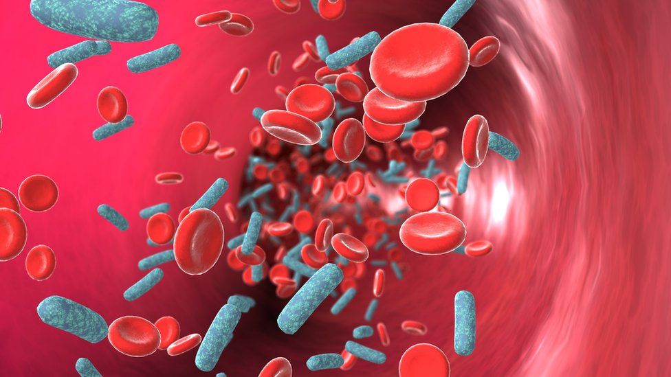
According to the WHO, there were nearly 49,000,000 cases of sepsis worldwide in 2017, and 11,000,000 of them were fatal. Compared to other more frequent causes of death, sepsis is a pathology that can often be effectively treated – if it is diagnosed in time. That is where Sepsiscan comes in – with a device that photographs skin and measures microcirculation of blood.
Monitoring of sepsis – micro and macro circulation
“Most infections are local in nature and affect one organ – as we are used to, for example, when we have a runny nose,” explains anesthesiologist-reanimatologist Sigita Kazūne. “Systemic symptoms usually do not occur. However, there is a possibility that the body’s response to the infection becomes so extreme that various distant tissues are damaged – often the kidneys, but also could be as bad as even the lungs or brain. That is the modern view of why sepsis occurs – a life-threatening pathology that can be caused by any type of infection (bacterial, viral or fungal) making the patient’s condition unstable and rapidly increasing the risk of mortality – by approximately 20%. However, there is a bright side – the current medical understanding of sepsis considers it one of the most preventable causes of death, with timely use of antibacterial therapy or surgical intervention. The key important thing to know is when a local infection turns into sepsis. In other words – we need better early diagnosis.”
At the beginning of the 21st century, new scientific publications showed that the early monitoring of blood microcirculation (large blood vessels) gives good results in the care of sepsis patients. That means – monitoring saturation of mixed venous blood, invasive blood pressure, and blood lactate level. However, current research and data suggests that the maximum benefit that can be obtained from these monitoring methods has been effectively reached. Therefore, the next step is to include microvascular (small blood vessels) methods in diagnostics – to analyze what is happening at the tissue level. Current methods capable of measuring that are either very simple and subjective (checking the recapillarization time with a stopwatch) or excessively complex (mucosal videomicroscopy – studying the flow of erythrocytes in the capillaries of the oral mucosa).
One of the early signs of sepsis (especially for patients in very serious cases) is a skin change called marbling. The color of the skin becomes bluish-violet, usually begins to develop from the knee area and in extreme cases it spreads to the whole body. If this symptom could be noticed at the very beginning and turned into a measurable quantity – observing the changes and how far they spread before they are even visible to the naked eye – then it would be possible to diagnose sepsis at a very early stage. Speaking in jargon, a technology is needed that is able to measure the oxygen content in the microcirculation bed, or how effective the oxygen supply to tissues is.

de Bruyne, Bernard et al. “Microvascular (Dys)Function and Clinical Outcome in Stable Coronary Disease.” Journal of the American College of Cardiology 67 10 (2016): 1170-1172 .
University spin-off
In 2017, S. Kazūne, together with the Institute of Atomic Physics and Spectroscopy of the University of Latvia (LU IAPS), started working on a project with the goal of using spectroscopic methods for monitoring sepsis patients. In the beginning, it was a small submission to the LU Foundation – the development of an optical non-invasive hybrid method for the diagnosis of sepsis – which successfully received funding, enabling the start of the first measurements with a customized hyperspectral camera. The results were promising and gave the project team the opportunity to write an application for funding from the European Regional Fund – obtaining enough funds to be able to collect additional data, perform analyses, process the results, and purchase the necessary parts for improving and adapting the equipment specifically for this purpose – allowing the creation of the first prototype device.
When the project was mature enough, the next step was commercializing the technology. S. Kazūne works in the Department of Anesthesiology of the Hospital of Traumatology and Orthopaedics, is an assistant professor at the Department of Anaesthesiology and Intensive Care at RSU, and also continues to participate in the LU IAPS projects, therefore she emphasizes: “My specialty is clinical medicine, and entrepreneurship is neither a familiar field nor, with three jobs, is there time to learn it.”
At the beginning of 2023, LU announced an auction for the right for businessmen to obtain a license to further develop this technology and commercialize it. Fortunately, the project didn’t have to go to a stranger. Didzis Rūtītis and Matīss Lācis worked in the university team for two years, and decided to continue with the project, winning the auction and founding the startup Sepsiscan.
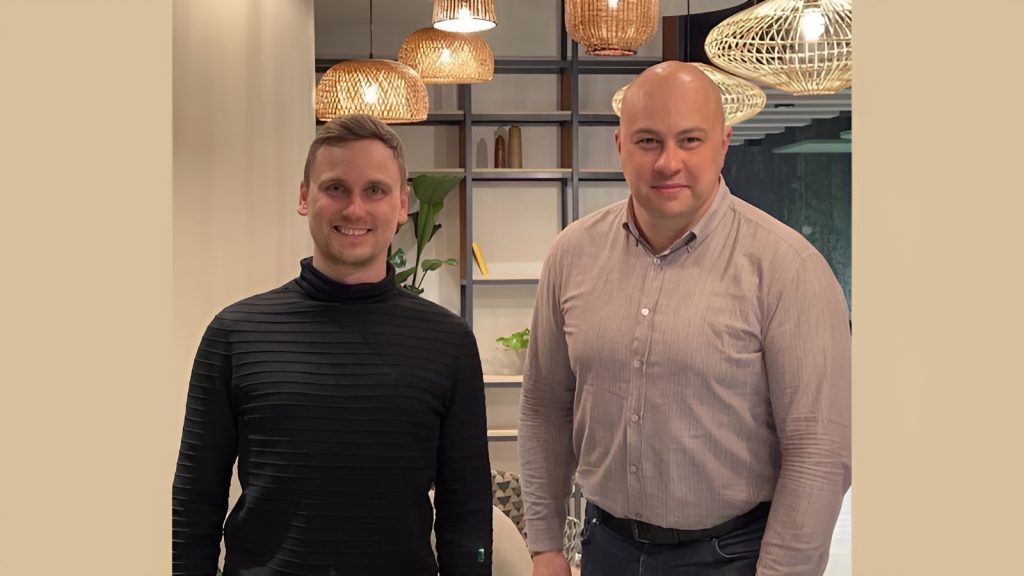
From the right – Matīss Lācis, Sepsiscan CTO, and Didzis Rūtītis, Sepsiscan CEO
The third prototype
«Our current goal is to complete a device prototype that is miniaturized for practical use. It is necessary both quite simply for commercialization purposes (so that it is easy to use and looks good), but also to be able to carry out a large-scale clinical study, which is necessary to register the device as a class 2A medical technology,” says Didzis, CEO of Sepsiscan.
It’s been only a year, but the company has made significant strides – completing the Estonia-based Health Founders accelerator and receiving first investments from Commercialization Reactor Fund. These vital funds are needed not only for technology development, but also for participation in industry congresses, innovation exhibitions and other opportunities for a new startup. It is difficult for medical technology companies to attract investments in the initial stages, because most investors want fast growth and profits, but the introduction of any innovations in medicine is a slow process. “In parallel with the development of the prototype, we are validating the idea and communicating with European hospitals and opinion leaders on the topic of microcirculation, who could support us with knowledge and networking opportunities. Since it is not easy to attract investments from traditional startup investors, we are looking for cooperation opportunities with pharmaceutical companies that are developing sepsis medication,” Didzis explains.
The company is currently working on the third version of the prototype. The first was tested in a hospital with a group of 100 patients – it could not be called a full-fledged clinical trial, as it was more of a technology pilot. However, the obtained data were interesting and promising, as a result of which they were used in the development of four publications. The second prototype was tested only within the university and is currently being improved and miniaturized to become the third, now commercial prototype, which will then also be used in a large-scale clinical trial.
“We have consulted with various external service providers who could help us prepare and conduct clinical studies, as well as obtain an ISO13485 certificate that confirms patient safety. We hope to conduct the research internationally – of course, it is easier to talk to hospitals here in Latvia, especially if they were already involved in the initial trials of the device, but at the same time, testing the device in other countries opens the door to the markets of those regions and understanding of their healthcare systems and legislative peculiarities,” says Didzis. “Finally, Europe has various innovation support programs that help deeptech and medical technology startups. Not only financially, but also with contacts – opening doors to cooperation with top hospitals.”
In addition to the need to conduct research outside of Latvia, CTO of Sepsiscan Matīss explains: “More extensive data is needed – with different kinds of people – microcirculation can affect life in different climate conditions, and the operation of the camera can be affected by different skin pigmentation.” S. Kazūne also emphasizes that in order to convince specialists outside of Latvia that this equipment is able to perform accurate measurements and help in the work with sepsis patients, it is necessary to obtain data from multicenter studies.

Sepsiscan device in use
Importance of software
Sepsiscan device functions as a very sensitive camera which is able to take an image of the subcutaneous tissue – depicting the microcirculation, which is not really visible to the naked eye. However, this is not enough – a special algorithm is also needed to process this acquired visual information. “We try to find an optimal balance between the accuracy of the equipment and fast, convenient operation,” says Matīss. “For example, the doctor should not have to hold the device perfectly still for an unreasonably long time. Additionally, an easy-to-use digital interface is important, which is why software development plays just as much a role as the device itself. In fact, there are currently no major changes planned for the device itself, a large part of the work is directly with the software.»
The startup’s planned business model would be to combine the sale of the device to hospitals with a software subscription model that would allow it to be continuously improved. There is also great potential in the future to start using machine learning, or so-called artificial intelligence, in the device’s operation process. However, Matīss emphasizes that it requires a lot of data – which would only be possible when the device is being actively and widely used. The data would be anonymized, of course, and the more there was, the more machine learning would help Sepsiscan become more accurate in processing the images.
Currently, the value of the device is theoretically more in monitoring patients – human bodies can be very different, and the base level of microcirculation can vary. It can be measured when the patient arrives at the hospital and then monitored regularly. However, machine learning would enable the doctor to immediately get a better idea of the patient’s microcirculation condition.
Opportunities and obstacles
In the view of S. Kazūne, the main advantages of Sepsican are – the possibility to quickly (no longer than measuring temperature) and non-invasively obtain data on the condition of the sepsis patient; to allow emergency departments to better decide where it is better to hospitalize the patient – in one of the clinical departments or in the intensive care unit; as well as the specificity of the method. For example, both blood lactate analysis and Sepsiscan actually measures the same thing – microcirculatory disturbances. But a high lactate level may not be the cause of sepsis, while poor oxygen saturation in the microcirculation blood vessels in the skin is much more specific for sepsis patients.
It is also important that such a new diagnostic method should be accompanied by corresponding changes in therapy. “Today, the treatment of sepsis is based on well-defined macrohemodynamic goals,” emphasizes S. Kazūne. “Howver, if we start to analyze microcirculation, there is a lack of research on what should be aimed for in therapy – that is, we need data that proves that when certain goals are reached, the patient’s health has improved. Until then, it is difficult to motivate hospitals and doctors to change the current guidelines. The clearer the therapeutic goals, the greater the demand for this type of equipment.»
From a technical point of view, Matīss mentions that a potential complication in the future is the chain of purchase of equipment parts: «We are evaluating different sources of parts, because we do not want to depend on one supplier. It is important to find the right cooperation partners – especially when we enter the market and need to produce a 1000 machines.»
However, in general, the founders of the company are optimistic: «On our side is the fact that the current medical view of sepsis is to measure microcirculation. How to do it? There is no consensus or common solution yet. We are at the right time and place to enter the market. But it won’t be fast – about a year to complete the development phase, the next for the clinical study, then at least a year for the registration of the device and only then – on to the market.»
This article was created in partnership with Medicus Bonus – a Latvian magazine for healthcare professionals – in a new series about healthcare innovation happening in Latvia. If you are a doctor, the subscription is free – just write to redakcija@medicusbonus.lv !